Translate

DO YOU KNOW?-3

DO YOU KNOW?-3
CREATININE CHEMISTRY

Translate

Wednesday 8 January 2020

KIDNEY DISEASES-3-CHRONIC KIDNEY DAMAGE(CKD)

CHRONIC KIDNEY DISEASE (CKD) &END STAGE KIDNEY DAMAGE(ESKD)

Fig-1

Chronic Kidney Damage or CKD can be defined as the step by step or slow loss of kidney functions over a long time period of months or years.
The main culprits for this CKD are generally High Blood Pressure(HBP) and Diabetes Mellitus(DM)
.Both uncontrolled high B.P and high sugar can damage the kidneys as follows:-
High B.P. would reduce the amount of blood flow to the kidney and thereby the kidney becomes undernourished.
Similarly high blood sugar will damage the glomerular filter system by sugar deposits. Thus the nephron can gradually lose its filtering capacity.
All causes of acute kidney failure are also fit for chronic kidney damages.

STAGES:-

1. Normal or above the normal stage- GFR (Glomerular Filter Rate)-Above 100 ml/min)
2.Mild stage -(GFR-60 to 99 ml/min)
3A.Moderate- (GFR-49 to 59 ml/min)
3B.Moderate- (GFR-35 to 49 mi/min)
4. Severe stage- (GFR- 19 to 29 ml/min)
5.End-stage   -  (GFR-<19 ml/min)
The first two stages can be reversed if attended in time with proper treatments. But they are asymptomatic and escape silently like thieves from our attention. Once the stage entered the third to fourth level the disease cannot be reversed as almost 50% of the kidney functions are destroyed.

Signs and Symptoms of CKD:-

1.Nausea and vomiting
2.Loss of hungry
3.Weakness and mental swings
4.Fatigue and tiredness
5. Frequent urination followed by reduced urination.
6.Foamy and bloody urine
7.Shortness of breath due to fluid collection in the     lungs
8.Chest tightness and pain due to fluid collection in the heart.
9.High and uncontrolled B.P due to fluid retention.
10.Insomnia
11.Skin itching due to elevated wastes such as uric acid, urea, and phosphorus levels in the blood.
12.Muscle cramps.
Many of the above symptoms are not specifically due to CKD and a proper diagnosis should be done to confirm it.

Causes:-

1.Diabetes mellitus
2.Chronic High B.P.
3.Tubule Infections
4.Glomerular infections (Glomerulonephritis)
5.Vesicoureteral Reflux, a condition in which there is a reverse flow of urine back to the kidney.
6.Renal cysts
7.Prolonged urinary obstruction due to urethral pressure, and enlarged prostate.
8.Recurrent kidney infections.

Risk Factors:-

1.Old age
2.Smoking
3.Alcohol
4.Obesity
5.Uncontrolled cardiovascular diseases
6.Uncontrolled diabetes M 
7.Hereditary Kidney disease

Complications:-

Fig-2

Chronic kidney disease can affect almost all parts of the body due to the following complications.
1. Elevation of potassium levels can affect heart functions.
2. Elevation of phosphorus level can produce skin rashes.
3. Elevation of calcium level can produce kidney stones, and bone weakness and heart problems
4. Fluid retention can cause pulmonary and heart edema, ankles, and leg swellings, and high pressure.
5. Anemia prevails due to lack of erythropoietin production by the kidneys and thereby the production of RBC is reduced.
6.Reduced sexual drive, reduced fertility, and erectile dysfunctions.
7.CNS damages lead to mental health and other complications.
8. Damage to the immune system causes increased vulnerability of the body to infections.
9.Pericarditis-A condition in which more fluid collection in the pericardium, the upper covering of the heart. (See Fig-2) above.

The Concept of Anemia-Erythropoietin:-


Apart from functioning as an excretory organ kidney plays other important roles to maintain our body's health.
Blood needs regular synthesis of Red Blood Cells (RBC) every day to compensate for its loss on aging. Erythropoietin is an important catalyst to synthesize new RBCs by the bone marrow. Erythropoietin is secreted by the kidneys and liver. Liver secretion is predominant in childhood. Afterward kidney secretion of erythropoietin is predominant in adulthood. At  ESKD kidneys lose almost 90% of the ability to produce erythropoietin and hence anemia develops rapidly.
Erythropoietin is produced by the interstitial fibroblasts in close association with the peritubular capillaries and the proximal convoluted tubule See Fig-3 above)
1.EPO is released into the blood circulation from the kidneys.
2.EPO tells the bone marrow to synthesize and release RBC.
3.RBC is released into the circulation. (Fig-3)
The erythropoietin (EPO) production is increased in a low oxygen atmosphere to produce more RBC. People living in high altitudes are subjected to produce high EPO production by the kidney as the availability of oxygen is low there.

TESTS AND DIAGNOSES:-

1.EPO TEST;-
EPO test is not so important to confirm CKD since there are many other factors that cause the irregularities of the EPO level in the blood.
The high altitude where there is low oxygen density would increase the erythropoietin level in the blood.
Polycythemia (abnormal elevation of RBC in the blood due to bone marrow defects would lower the release of erythropoietin by the kidneys.
In the case of end-stage kidney damage (ESKD) the kidneys are fully disabled to produce EPO. Hence the EPO test may be a marker of the presence of ESKD.
The normal EPO level in the blood is 4 to 40 international units per liter (IU/L) of the blood.
Usually in kidneys the proximal convoluted tubule, the peritubular capillaries in the cortex are the areas where the interstitium produces EPO.
For an elaborate study click here
2. Diagnosis for ESKD involves a 24 hours urine test
3.Blood creatinine analysis:-Normal creatinine level is 0.7 to 1.2 mg/dl
4.Blood Urea Nitrogen tests:- Normal urea level is 14 to 25 mg/dl
The best marker to predict any type of kidney damage is the micro protein urea tests which we already described in previous chapters. To see that click  here
KIDNEY INJURY MOLECULE (KIM)TEST:-
The most reliable and accurate diagnostic test to predict AKD and CKD is the KIM-1 test.
KIM is a transmembrane glycoprotein present in the epithelial layer of the proximal tubule.Normal urine contains 0 to 0.7ng/ml.But in the injured kidney the level raises above 3.7 ng/ml which predicts kidney damage.
The test is more accurate to indicate the presence of AKD but can also be used as a reliable marker test to predict CKD.
Diet control, dialysis followed by EPO injections, or kidney transplants are life-saving formulas for those who reached end-stage kidney disease(ESKD).
ERYTHROPOIETIN INJECTIONS:-
These are human recombinant erythropoiesis-stimulating injections that must be given to the kidney patients either in dialysis or not.
The injections should be given at least three times per week and the dosage should be determined by the doctor.
Availability:-1)  2000 units/ml
                     2)  3000 units/ml
                     3)  4000 units/ml
                     4) 10000 units/ml
The initial treatment should be started when the hemoglobin count falls below 10 gram per deciliter of the blood.
As the count raises reduce the dosage accordingly.
Usual Dosage:-50 to 100 units per kg via IV or SC route.
Hemodialysis & Peritoneal dialysis will be presented as separate posts in the future.

Thursday 5 December 2019

KIDNEY DISEASE-2 ACUTE(AKD)

ACUTE KIDNEY FAILURE(AKF or AKD)

Acute kidney damage is a condition in which the kidneys are suddenly unable to clean your blood from harmful wastes, dangerous metabolites, and thereby let them accumulate in your body to a dangerous life-threatening level.
The condition develops in a short period of time in a few days or even in a few hours and can be fatal and need intensive hospital care.
Acute kidney failure or damage may sometimes be reversed to a normal condition if there are no other problems.
AKD may be confirmed when there is a 50 to >50% increase in serum creatinine levels.
Fig 1
     A sudden episode of kidney failure occurs for the following reasons.
1.Drugs of abuses like alcohol, narcotics, pain killers, antibiotics, and any poisonous drug whose metabolite can harm kidneys.
Alcohol over usage can cause kidney muscular necrosis as its metabolites such as acetaldehyde without further dehydration to acetic acid may leak from the liver and cause necrosis to the kidney tubular cells.
Over drink of alcohol may cause high B.P which is harmful to the kidney.
2.Pain Killer abuse. Non-Steroidal Anti Inflammatory Drugs can cause kidney damages if overdosed. They inhibit the production of important biological mediators such as prostaglandins. Prostaglandin PgE2 and Prostacyclin PgL2 are the important biological mediators that cause free renal blood flow by dilating the blood vessels and thereby facilitate and increase glomerular filtration.NSAIDs such as ibuprofen, naproxen, phenoxybenzamine, indomethacin, and aspirin are all inhibiting cyclooxygenase (COX-1 &2)and thereby inhibiting the prostaglandin synthesis.
3.Acute tubular necrosis. This condition can happen due to the consumption of foods, drinks, and drugs which can be converted into harmful excretory products or metabolites.
(e.g)Iodine contaminated contrast media for radio analysis.
After an intramuscular administration of iodinated contrast media, some patients reported within a few days of acute kidney injury developed which is
notified by an increase in creatinine and blood urea level above the normal.

ETIOLOGY

Fig 1(Table)
In the above Table, a few examples have been described for your reference.
1. Heavy consumption of ethanol can result in sudden acute renal injury in many ways.
The liver metabolizes ethanol into acetaldehyde by the enzyme alcohol dehydrogenase. Acetaldehyde is further metabolized by aldehyde dehydrogenase to harmless acetic acid which is decomposed to water and carbon dioxide.
But in some unhealthy situations of liver damage for example during hypothyroidism the liver may not properly metabolize acetaldehyde due to lack of aldehyde dehydrate. In this case the unmetabolized acetaldehyde can reach the kidneys and harm both kidneys as well as the liver. Remember acetaldehyde is highly carcinogenic.
Heavy consumption of alcohol can raise your B.P.and sugar in the blood and thereby reducing the blood supply to the kidneys and thereby injure them.
2. Methanol also produces dangerous metabolites like formaldehyde and formic acid both are harmful to the liver as well as to kidneys.
3. All opioids used as pain killers, diarrhea, and cough suppressants are all harmful to the kidneys.
Codeine phosphate, an opioid that is used in cough syrups as an antitussive (cough suppressant) agent can harm your kidney by its metabolites morphine, norcodeine, and codeine-6-phosphate.
4. Aminoglycosides such as neomycin, tobramycin, gentamicin, and streptomycin are all excreted by the kidney unchanged. The damage is mainly at the proximal tubule cells after passing through the glomerular filtration.
5. Heavy metals such as cadmium, chromium, and mercury are all excreted through the kidneys unchanged. Cadmium poisoning is very common in our life. Rice produced by using phosphate fertilizers, usage of plastics, pigments, and platings,
batteries, exposing to sewage sludge are all the possible man-made sources of cadmium poisoning. Cigarette smoking is one of the sources to get cadmium exposure to heavy smokers. Chronic exposure to cadmium may damage your kidney.
5. Pain killers such as ibuprofen(brufen), naproxen(Proxen), aspirin are harming the kidneys by inhibiting the synthesis of biological mediators prostaglandin, and prostacyclin.

Symptoms:-

The following are the symptoms of acute kidney failure.
1.Reduced urine output.Water retention and accumulation in the body.Swelling of the ankles, legs foot, and fingers.
2.Blood in the urine.
3.Tiredness and fatigue
4.Skin itching and necrosis
5.Cardiovascular diseases due to water accumulation inside the pericardial space.Arrhythmias.
6.Shortness of breath due to fluid accumulation in the lungs.
7.Nausea
8.Mood disturbances
9.Weakness.
10.Seizures and coma.

DIAGNOSIS:-

1.A.URINARY EVALUATION:-In AKD changes the blood creatinine level is not sensitive for early diagnosis.
Urinary levels of 
a)Kidney Injury Molecule-1(KIM-1)
b)N-acetyl-β-D-glucosaminidase(NAG)
c)matrix metalloproteinase-9(MMP-9)
are evaluated as biomarkers for the detection of AKD.
AKD is often seen in the setting of multiple organs failure and sepsis. 
The top and noninvasive biomarker is the KIM-1 biomarker. This is the more common biomarker for the early detection of AKD. This is a transmembrane protein. This is not detectable in normal kidney tissue or urine. But in dedifferentiated (PCT)epithelial cells and urine after an ischemic or toxic injury and in renal cancer this biomarker is expressed at a high level.
The NAG biomarker takes the next important biomarker place.
The least sensitive is the result of the MMP-9 biomarker.
The absence of sensitive biomarkers the diagnosis for early detection of AKD has had impaired and detrimental results. These biomarkers are micro molecules should be absent in the urine in normal persons
Urine tests to assess kidney diseases are usually performed with 24 hours of urine collection. To collect the sample the first urine which empties the bladder should be discarded and the next 24-hour urine should be collected for the tests.
1.B.Urinary Minerals Level.:-
The normal urine potassium level is 25 to 125 milliequivalent per liter. (mEq/L). But in case of AKD, this level goes down to below 25 mEq/L.A. Serum potassium test should be done to confirm the rise in potassium level above the normal limit(3.6 to 5.2 mmol/L)
(1mEq of potassium=39mg)
(To convert mmol/L to mg/dL multiply the figure  by 18
To convert mg/dL to mmol/L, divide the figure by 18)
Similarly calcium in the urine (100 to 300 mg/day)should be done.
Urine magnesium should be less than 24 mg in a 24-hour urine collection.
Similarly, phosphorus should be measured in a 24 urine collection
2.Serum Creatinine Tests:
If the serum creatinine levels are above 3mg per deciliter then it could be assessed as there are epithelial damages and dedifferentiation in tubular tissues through which the filtered creatinine is leaked passively back into the blood. Another reason is if there are inflammations in the Bowman's capsules as in the case of glomerulonephritis which results in improper creatinine and urea filtration.
3.Blood Urea Nitrogen (BUN):-
The normal serum BUN level is between 7 to 25 mg per deciliter of the blood. In the case of AKD and CKD this may elevate beyond 30 mg/dL.
CORRECTIONS
Kidney damage in a short period can some times be reverted to normal if it is due to the following causes.
1.If the condition which slows the blood flow to the kidneys is solved. For example, blood and fluid loss. This can be corrected by proper blood transfusion and fluid infusion.
2.Discontinuation of the usage of painkillers
3.Immediate care of heart and liver diseases
4. Change B.P medication from ARB inhibitors (Losartan) or ACE inhibitors (Enalapril)to any other alternative medicine as ARB, and ACEI disturbs the aldosterone, vasopressin and adrenergic pathways and thereby harm kidney functions.
5.Urinary infection if any should be treated in time.
6. Severe dehydration due to diarrhea or vomiting, loss of fluid should be corrected immediately by drinks of infusion. The loss of electrolytes should be compensated.
7. Anaphylaxis and other allergic conditions should be cared for in time.
8. Avoid alcohol, tobacco, opioids, cocaine, like drugs of abuse.
9. Avoid amphetamines.
10. Take care of your prostate.
11. In sudden hemorrhages or blood loss due to accidents blood should be immediately compensated by infusion.
Continued...


Saturday 30 November 2019

KIDNEYS (RENALS) FAILURE-1 INTRODUCTORY

A SHORT INTRODUCTION TO KIDNEYS(RENTALS) -1

Fig-1

Kidneys are two bean-shaped excretory organs located against the back muscles in the upper abdominal area. They sit opposite each other at the right and left sides of the spine. The right kidney is fitted a little lower than the left kidney to accommodate the liver. See Fig-1 above.

Blood Supply To Kidneys(The Renal Circulation):-

Fig 2
Despite their smaller sizes the kidneys receive almost 20% of the total cardiac output. They are well supplied with renal arteries which branch from the aorta at the abdominal region. Kidneys after receiving about 20 to 25% of the cardiac output it filters and cleans the blood from many harmful wastes and impurities such as urea, creatinine, hippurates, uric acid, and many metabolic medical wastes. Also kidneys excrete excess minerals to keep the balance of the electrolytes in the body. After cleaning the blood they consume 3% of the blood for their own oxygen need. The remaining cleaned and deoxygenated blood is drained into the veins and return back to the heart. (See Fig.2 above).

THE NEPHRONS AND THE URINE FORMATION:-

Fig-3 A

Fig 3B
The two figures are shown above (3A and 3B)are for illustrative purposes only. The nephrons are the fundamental units of the kidneys. There are about one million nephrons in each kidney.
These number of nephrons are really the Grace and Gift of the Almighty to the human lives. In our everyday life, we are abusing our kidneys. By our unhealthy eating and drinking habits, stress situations, the dirty atmosphere we are killing the nephrons smoothly. To combat these abuses God has given us this huge number of nephrons with a Great Kindness. All praises to the Almighty God.
As shown in the figures above a nephron is composed of the following parts.
1.The Bowman's Capsule:
The cup-shaped head like part. This is the first part of the nephron. It is like a sac enclosing the glomerulus. The sac is 8 to 10 nm. The blood that enters into this portion is already ultra-filtered by leaving back the macrocells and proteins. The filtrate contains 0.05 % of proteins.
2.Glomerulus:-
A small ball-shaped knitted form of tiny capillaries fitted inside the Bowman's capsule. Blood is filtered in this portion of the nephron.
3.Proximal Convoluted Tubule:-
Pre or Proximal Convoluted Tubule (PCT) is the coiled tubule immediately following down the Bowman's Capsule. In this part when the filtrate passes, important nutrients like glucose, and proteins in the form of peptides are reabsorbed back into the blood. Water is absorbed along with sodium and bicarbonate ions in this part. Further in this region urea, chloride, potassium, lactate, phosphate, and bicarbonate are also absorbed back into the blood.
4.Loop Of Henley (Descending):-
This is the narrowed descending portion of the PCT.
After some water is absorbed along with various ions mentioned above in the PCT a one-third of the filtrate which is hypotonic reaches in this portion. Here the tubule is highly permeable to water only and not the solute. Water is reabsorbed back to the blood by leaving a highly concentrated filtrate to pass down into the bottom of the loop. Afterward the hypertonic filtrate passes upwards to the ascending loop of Henley.
5.Loop of Henley (Ascending):-
In this portion the loop cells are permeable only o the solutes and not to the water. Thus solutes like sodium, potassium, and chloride ions are reabsorbed back to the blood, with the exchange of hydrogen out into the filtrate. Hence the filtrate is more and more diluted to hypotonic with water.
In general the movement of the luminal fluid in the loop region is sufficiently slow in order to maintain the concentration of the urine to keep the osmolarity balance in the medulla. Sodium, potassium, calcium, chloride, and bicarbonate ions are reabsorbed in the ascending loop of Henley.
6.Distal Convoluted Tubule (DCT):-
This portion of the nephron is located again back upward to the cortex region near the end part of the nephron. After passing through the ascending loop the filtrate enters into the lumen of the DCT. Here minerals like calcium, sodium, and chloride are reabsorbed with the exchange of potassium by the action of vasopressin (Anti Diuretic Hormone)  secreted from the posterior pituitary. The hydrogen ion is also secreted into the filtrate in order to maintain the pH of the urine around 5(acidic).
7. Collecting Tubules And Collecting Ducts:-
Collecting tubule is the nonvascularized portion of the DCT which drains the urine into the collecting duct. All the collecting ducts from all the nephrons are combined to form a single ureter which descends down and open at the upper mouth of the bladder.

Ureter:-

Ureters are two single tubes from each kidney receives urine from the collecting ducts and carries urine down to the urinary bladder.
THE LOCATION OF THE NEPHRON
Fig 4 A


Fig 4 B

The location of the nephrons are simplified diagrammatically in the above two figures (Fig 4A and 4B)
A single nephron has been represented for a simple understanding of their locations inside the kidney. A rough cross-section of the kidney has been figured above. In the cross-section (4B) the nephron except for its loop part the remaining parts like Bowman's capsule, the coiled tubules are all situated at the peripheral cortex of the kidney. Only the loop of Henley and the collecting ducts are situated in the medullary region.
In the cross-section (Fig 4A) it has been shown that the cortex is more vascularised than the medulla. Only the loop portion is vascularised in the medulla. The collecting tubule and the collecting duct are exempted from vascularization as there are no cleansing activities there.
one-third of the medullary region is occupied by the pelvis. All collecting ducts from various nephrons are grouped into the pelvis. All pelvises are unified to form the single ureter. This is the simplified physiology and anatomy of the kidney.                           Continued......


Thursday 21 November 2019

THYROID UNDER ACTIVITY EFFECTS IN ECG

ECG EFFECTS OF HYPOTHYROIDISM

HYPOTHYROIDISM EFFECTS

https://www.linkedin.com/pulse/thyroid-under-activity-ecg-seyed-ali/?trackingId=8sJR43x6QnKqcMkADkNKWg%3D%3D
Hypothyroidism is the condition in which the thyroid gland is unable to secrete sufficient quantity of its hormone thyroxine (T4). This disease is due to various causes such as 1.thyroid inflammation or thyroid cell necrosis due to a disease called Hashimoto Autoimmunity Disease.
This is one of the very common and very important causes of thyroid dysfunction.
Our body is equipped by nature with self-protection forces against invading pathogenic cells such as bacteria. When an invading pathogen enters into our body, our immune apparatus immediately responds to it by sending fighting cells to the area of invasion to kill the pathogen.
But due to some unexplained situation the fighting cells confused with healthy cells such as cells of the thyroid gland and kill them. This is known as Autoimmune Disease, or Hashimoto's Disease.
2.Medications for Hyperthyroidism such as Propylthiouracil, and Methimazole(Tapazole), Potassium or Sodium Iodide, and Radio Iodine (I131) if overdosed may cause thyroid gland damage.
3.Insufficient secretion of TSH from the pituitary due to some unknown pituitary disease
4.Thyroidectomy-Removal of the gland
4.Insufficient iodine intake.
To go further is beyond the scope of this ECG article. Hence to study further regarding causes, symptoms, and treatment please click here.

Hypothyroidism in ECG

In the above figure the effects of Hypothyroidism are apparent in examining the ECG.
In the lead-II recording, one can observe the elongated R-R interval which represents the presence of bradycardia (slower heart rate)
The absence of P-wave and the elongated P-R segment indicates the dysfunction of the atria.
Depression of ST-segment and the deformation of T-wave indicates the dysfunctions of the ventricles and the presence of angina.
Conclusively untreated long term hypothyroidism can precipitate many serious cardiac issues.

Monday 11 November 2019

ECG CHANGES IN THYROID OVERACTIVITY

ECG CHANGES DURING HYPERTHYROIDISM

Fig-1
Hyperthyroidism is a serious condition in which the thyroid gland becomes overactive and produces many complications.
The thyroid is a tiny butterfly-shaped gland that is located in front of the neck just below Adam's apple.
It is producing the hormone thyroxine (T4) and triiodothyronine T3 releasing them in circulation.T3 (Triiodothyronine) which is more active than T4 (tetraiodothyronine) is highly unstable and is soon converted into T4. For details click here.
Due to the following causes hyperthyroidism may occur.
1.Excessive intake of thyroid tablets
2.Excessive secretion of TSH (Thyroid     Stimulating Hormone)
3.Grave's Disease.An autoimmune disease in which the thyroid gland produces more hormone.
4.Inflammations of the thyroid gland (Thyroiditis)
5.High iodine intake
6.Functioning adenoma
7.Severe Multinodular Goiter(SMNG)
Normally the body uses thyroid hormone to speed up metabolism, which raises and maintains the body temperature within limits.
But if the thyroid secretes high levels of the hormone then the temperature raises more rapidly with dangerous levels of anabolism and catabolism which raises energy level beyond the control. Blood pressure raises, with increased heart rate and many more complications.
Symptoms:-
1.Tremor, exciting, nervousness.
2.Anxiety
3.Tiredness, and weakness
4.Mood swings
5.High temperatures(Hyperthermia)
6.Sudden unexpected weight loss
7.Goiter(Swollen neck)
6.Increased heartbeats (Tachyarrhythmia), palpitations.
7.Rapid bowel movements
8.Sleeplessness
9.Thinning and brittle skin and hair.
10.Disturbed menstrual cycles.
Thyroid Storm:-
If hyperthyroidism is not properly cared for and treated in time it will lead to this acute condition in which all symptoms mentioned above will rise to a life-threatening dangerous level with fatal results. 
ECG EFFECTS;-
Fig-2A
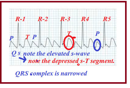
In the above Fig-2A a model ECG taken during Hyperthyroidism has been present for reference.
Observe the waves P, Q, R, S, and T.The normal rhythm is highly disturbed as we have seen in past articles. To get a detailed description of a normal sinus rhythm please click here.
Fig-2B
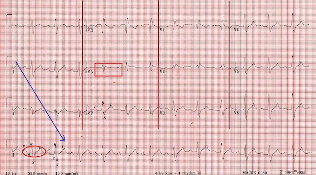
In the above Fig-2A and Fig 2B note the records by Leads-II, III, I aVL, and aVF which give the picture of rapid heartbeats(tachyarrhythmias) by the narrowed QRS complex and the shortened R-R intervals.
Short P-R intervals indicate the rapid contractions of the atria followed by the immediate ventricular responses.
The depression of S-wave followed by elevation of the ST segment indicates the strain on the ventricles. These changes are predicting a heart attack to follow ventricular fibrillation.
Conclusively thyroid overactivity and thyroid storm should be treated in time to avoid fatal consequences.
                               (Next: Thyroid Under Activity)


Monday 4 November 2019

EFFECTS OF MEDICINES IN ECG-BETA BLOCKERS AND CALCIUM CHANNEL BLOCKERS.-3

1.ATENOLOL(TENORMIN), PROPRANOLOL(INDERAL) AND CALCIUM CHANNEL BLOCKERS(CCB)

Atenolol is a B.P medicine commonly available in the pharmacy under the brand name as Tenormin.
It is technically known under the group as a Beta Receptor Blocker or simply as Beta Blocker.
Briefly we can say that beta-blockers are drugs that lower the blood pressure by lowering the heart rate by blocking beta-1 receptors of the adrenergic nervous system which is more concentrated on the cardiovascular network. The result also included dilation of the blood vessels and reduction of the heart rate.
This drug has notable effects on the ECG if overdosed.
Fig-1


A normal heart rhythm has been shown diagrammatically in Fig-1 above. In that, the waves P, Q, R, S, and T are clear and distinguishable.
Atenolol is a safe B.P medicine with a wider therapeutic index. It is highly water-soluble and is eliminated in the urine. It is less fat-soluble and hence it is less likely to pass into the blood-brain barrier to produce CNS effects.
But as with any other medicine if overdosed or misused by otherwise a normal person atenolol propranolol and any other beta-blocker will reduce heart rate, blood pressure followed by fatal CHF. This can be noticed in the ECG by prolonged PR segment, wider QRS complex, and elevated or depressed ST segment.
Fig-2
In Fig 2 above a model, ECG has been shown with the effects of all beta-blockers such as atenolol, propranolol(Inderal), metoprolol(Lopressor)and calcium channel blockers such as diltiazem(Dilzem, Cartia), verapamil(Isoptin), amlodipine(Norvasc) and nifedipine(Procardia).
In the above ECG examine the lead-II the master lead record which shows the depressed ST segment, with a more depressed S-wave, and the absence of Q-wave.
The aVL record shows (marked by a small red squire) the absence of P, and T-wave and elongated P-R and S-T segments or intervals.
This indicates severe bradycardia (reduced heartbeat)
extremely low pressure and congestive heart failure (CHF)
                                                                                                   
                                                                                                         Fig 3A
Fig 3B
In the above two diagrams (Fig 3A and 3B) the effects of verapamil (Isoptin) a CCB drug on ECG taken in an old man who has been overdosed by the drug.
In Fig 3A a simulated image has been explained.
Note down the marked deviation of Phase-0 more towards the right. This is because calcium channels are blocked and the entry of calcium is restricted with the result of the shortening of the plateau Phase-2.
The deviation has been explained in the down by elongated PR-interval widening of the QRS complex and the elongated ST-segment.
These changes can be easily noted in Fig-3B in the Lead-II records. The elongated intervals and segments and the QRS widening are marked by a blue rectangle.
Conclusively the ECG effects of both beta-blockers and CCBs are similar by many factors such as elongated PR-intervals, widened QRS-complex, elongated ST-segment, etc which indicates sinus bradycardia followed by CHF.
Thank You


Friday 18 October 2019

ECG IN HYPERKALEMIA

HIGH POTASSIUM LEVEL IN ECG

Hyperkalemia is a harsh condition in which blood potassium levels are above normal in the blood. This is one of the common conditions during End-Stage Kidney Disease(ESKD). But due to some other causes also this condition may occur.
For example frequent uses of Trimethoprim-Sulfamethoxazole combination (Septrin, Septra, and Bactrim)may raise potassium level in the blood. Beware of these antibiotics if the patient is aged above 60 years.
Here we are about to see only how this condition is reflected in the ECG.
Fig-1
In the above Fig-1 a normal ECG has been shown. In that one can observe a normal sinus rhythm with a clear P, Q, R, S, and T-waves.
In the following Fig-2A and 2B each, an ECG has been taken during the condition of high potassium level in the blood.
Fig-2A


Fig 2B


In general hyperkalemia causes the reduction in heartbeats or frequency of contraction(cardiac chronotropic) and increasing the force of contractility (cardiac inotropy). It causes errors in cell polarization and depolarization. Irregular beats.Cardiac arrhythmias.Bradycardia or slowing of heartbeats. If left uncorrected it may result in cardiac arrest.

In the above Fig-2A, the precordial leads V3 and V4 show the stages of abnormalities develops in the sinus rhythm due to the high level of potassium in the blood.
The first stage shows normal P-wave with the abnormal decay of the QRS complex followed by the elevated T-wave.
The second stage shows the flattening of P-wave and the third stage shows the total eclipse of P-wave.
The QRS complex is shortened and narrowed.
T-wave is high peaked.
Segment-wise the PR-seg.is elongated and the ST-seg.is elevated to predict the event of a heart attack.
The P -wave disappearance is due to the atrial nonresponse to the SA node triggering.
The QRS shortening and narrowing indicate and predict ventricular fibrillation.
The elevated T-wave indicates the delay in ventricular repolarization.
Control potassium intake by avoiding potassium-rich foods such as unpeeled fruits, red meats, raisins, prunes, potatoes, and drinks like tender coconut, orange, banana, grapefruit, tomato, prune and apricot juices.
Symptoms of hyperkalemia are mild or asymptomatic at stage one 
but shows fatigue, tiredness, tingling and numbness, nausea and vomiting, breathing trouble, and chest pain.


BRAIN MAPPING

BRAIN MEANDERING PATHWAY                                                                         Maturity, the thinking goes, comes with age...