Translate

DO YOU KNOW?-3

DO YOU KNOW?-3
CREATININE CHEMISTRY

Translate

Monday 11 November 2019

ECG CHANGES IN THYROID OVERACTIVITY

ECG CHANGES DURING HYPERTHYROIDISM

Fig-1
Hyperthyroidism is a serious condition in which the thyroid gland becomes overactive and produces many complications.
The thyroid is a tiny butterfly-shaped gland that is located in front of the neck just below Adam's apple.
It is producing the hormone thyroxine (T4) and triiodothyronine T3 releasing them in circulation.T3 (Triiodothyronine) which is more active than T4 (tetraiodothyronine) is highly unstable and is soon converted into T4. For details click here.
Due to the following causes hyperthyroidism may occur.
1.Excessive intake of thyroid tablets
2.Excessive secretion of TSH (Thyroid     Stimulating Hormone)
3.Grave's Disease.An autoimmune disease in which the thyroid gland produces more hormone.
4.Inflammations of the thyroid gland (Thyroiditis)
5.High iodine intake
6.Functioning adenoma
7.Severe Multinodular Goiter(SMNG)
Normally the body uses thyroid hormone to speed up metabolism, which raises and maintains the body temperature within limits.
But if the thyroid secretes high levels of the hormone then the temperature raises more rapidly with dangerous levels of anabolism and catabolism which raises energy level beyond the control. Blood pressure raises, with increased heart rate and many more complications.
Symptoms:-
1.Tremor, exciting, nervousness.
2.Anxiety
3.Tiredness, and weakness
4.Mood swings
5.High temperatures(Hyperthermia)
6.Sudden unexpected weight loss
7.Goiter(Swollen neck)
6.Increased heartbeats (Tachyarrhythmia), palpitations.
7.Rapid bowel movements
8.Sleeplessness
9.Thinning and brittle skin and hair.
10.Disturbed menstrual cycles.
Thyroid Storm:-
If hyperthyroidism is not properly cared for and treated in time it will lead to this acute condition in which all symptoms mentioned above will rise to a life-threatening dangerous level with fatal results. 
ECG EFFECTS;-
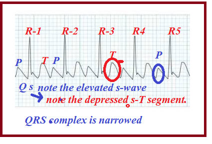
Fig-2A
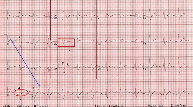
In the above Fig-2A a model ECG taken during Hyperthyroidism has been present for reference.
Observe the waves P, Q, R, S, and T.The normal rhythm is highly disturbed as we have seen in past articles. To get a detailed description of a normal sinus rhythm please click here.
Fig-2B
In the above Fig-2A and Fig 2B note the records by Leads-II, III, I aVL, and aVF which give the picture of rapid heartbeats(tachyarrhythmias) by the narrowed QRS complex and the shortened R-R intervals.
Short P-R intervals indicate the rapid contractions of the atria followed by the immediate ventricular responses.
The depression of S-wave followed by elevation of the ST segment indicates the strain on the ventricles. These changes are predicting a heart attack to follow ventricular fibrillation.
Conclusively thyroid overactivity and thyroid storm should be treated in time to avoid fatal consequences.
                               (Next: Thyroid Under Activity)


Monday 4 November 2019

EFFECTS OF MEDICINES IN ECG-BETA BLOCKERS AND CALCIUM CHANNEL BLOCKERS.-3

1.ATENOLOL(TENORMIN), PROPRANOLOL(INDERAL) AND CALCIUM CHANNEL BLOCKERS(CCB)

Atenolol is a B.P medicine commonly available in the pharmacy under the brand name as Tenormin.
It is technically known under the group as a Beta Receptor Blocker or simply as Beta Blocker.
Briefly we can say that beta-blockers are drugs that lower the blood pressure by lowering the heart rate by blocking beta-1 receptors of the adrenergic nervous system which is more concentrated on the cardiovascular network. The result also included dilation of the blood vessels and reduction of the heart rate.
This drug has notable effects on the ECG if overdosed.
Fig-1


A normal heart rhythm has been shown diagrammatically in Fig-1 above. In that, the waves P, Q, R, S, and T are clear and distinguishable.
Atenolol is a safe B.P medicine with a wider therapeutic index. It is highly water-soluble and is eliminated in the urine. It is less fat-soluble and hence it is less likely to pass into the blood-brain barrier to produce CNS effects.
But as with any other medicine if overdosed or misused by otherwise a normal person atenolol propranolol and any other beta-blocker will reduce heart rate, blood pressure followed by fatal CHF. This can be noticed in the ECG by prolonged PR segment, wider QRS complex, and elevated or depressed ST segment.
Fig-2
In Fig 2 above a model, ECG has been shown with the effects of all beta-blockers such as atenolol, propranolol(Inderal), metoprolol(Lopressor)and calcium channel blockers such as diltiazem(Dilzem, Cartia), verapamil(Isoptin), amlodipine(Norvasc) and nifedipine(Procardia).
In the above ECG examine the lead-II the master lead record which shows the depressed ST segment, with a more depressed S-wave, and the absence of Q-wave.
The aVL record shows (marked by a small red squire) the absence of P, and T-wave and elongated P-R and S-T segments or intervals.
This indicates severe bradycardia (reduced heartbeat)
extremely low pressure and congestive heart failure (CHF)
                                                                                                   
                                                                                                         Fig 3A
Fig 3B
In the above two diagrams (Fig 3A and 3B) the effects of verapamil (Isoptin) a CCB drug on ECG taken in an old man who has been overdosed by the drug.
In Fig 3A a simulated image has been explained.
Note down the marked deviation of Phase-0 more towards the right. This is because calcium channels are blocked and the entry of calcium is restricted with the result of the shortening of the plateau Phase-2.
The deviation has been explained in the down by elongated PR-interval widening of the QRS complex and the elongated ST-segment.
These changes can be easily noted in Fig-3B in the Lead-II records. The elongated intervals and segments and the QRS widening are marked by a blue rectangle.
Conclusively the ECG effects of both beta-blockers and CCBs are similar by many factors such as elongated PR-intervals, widened QRS-complex, elongated ST-segment, etc which indicates sinus bradycardia followed by CHF.
Thank You


Friday 18 October 2019

ECG IN HYPERKALEMIA

HIGH POTASSIUM LEVEL IN ECG

Hyperkalemia is a harsh condition in which blood potassium levels are above normal in the blood. This is one of the common conditions during End-Stage Kidney Disease(ESKD). But due to some other causes also this condition may occur.
For example frequent uses of Trimethoprim-Sulfamethoxazole combination (Septrin, Septra, and Bactrim)may raise potassium level in the blood. Beware of these antibiotics if the patient is aged above 60 years.
Here we are about to see only how this condition is reflected in the ECG.
Fig-1
In the above Fig-1 a normal ECG has been shown. In that one can observe a normal sinus rhythm with a clear P, Q, R, S, and T-waves.
In the following Fig-2A and 2B each, an ECG has been taken during the condition of high potassium level in the blood.
Fig-2A


Fig 2B


In general hyperkalemia causes the reduction in heartbeats or frequency of contraction(cardiac chronotropic) and increasing the force of contractility (cardiac inotropy). It causes errors in cell polarization and depolarization. Irregular beats.Cardiac arrhythmias.Bradycardia or slowing of heartbeats. If left uncorrected it may result in cardiac arrest.

In the above Fig-2A, the precordial leads V3 and V4 show the stages of abnormalities develops in the sinus rhythm due to the high level of potassium in the blood.
The first stage shows normal P-wave with the abnormal decay of the QRS complex followed by the elevated T-wave.
The second stage shows the flattening of P-wave and the third stage shows the total eclipse of P-wave.
The QRS complex is shortened and narrowed.
T-wave is high peaked.
Segment-wise the PR-seg.is elongated and the ST-seg.is elevated to predict the event of a heart attack.
The P -wave disappearance is due to the atrial nonresponse to the SA node triggering.
The QRS shortening and narrowing indicate and predict ventricular fibrillation.
The elevated T-wave indicates the delay in ventricular repolarization.
Control potassium intake by avoiding potassium-rich foods such as unpeeled fruits, red meats, raisins, prunes, potatoes, and drinks like tender coconut, orange, banana, grapefruit, tomato, prune and apricot juices.
Symptoms of hyperkalemia are mild or asymptomatic at stage one 
but shows fatigue, tiredness, tingling and numbness, nausea and vomiting, breathing trouble, and chest pain.


Sunday 6 October 2019

MEDICATIONS THAT AFFECTS YOUR ECG-2

TRYCYCLIC ANTIDEPRESSANTS OVERDOSE

Amitriptylin
Tricyclic antidepressants are prescribed by many doctors to treat depressions. They are classified into secondary amines and tertiary amines. Among this amitriptyline is one of the tertiary amines available in the market by the name of Tryptizol, Tryptomer, and Elavil, etc.
Many people are using this drug under any one of the above trade names for their depressive episodes. The following ECG image can reveal how this drug is affecting your heart.
Fig-1
The main effect of amitriptyline on the heart is QT prolongation and a fast heartbeat. See in Fig 1 above the QRS complex is widened in a manner to embed the T-wave so that the QT-interval is elevated and prolonged.P-Q interval also prolonged.
DOXEPIN:-

Fig-2

Doxepin is another tertiary amine very similar to amitriptyline. In Fig-2 the effect of doxepin is shown with a similar pattern of ECG changes to that of amitriptyline. Generally all tricyclic antidepressants produce similar patterns of ECG changes. They produce ventricular tachycardia with prolonged QT-interval.


Wednesday 2 October 2019

MEDICATIONS THAT AFFECTS YOUR ECG-1


DIGOXIN

Brand Name:Lanoxin,Digitek

Digoxin is the drug that is used frequently to patients suffering from Congestive Heart Failure.
Digitalis Lanata or purpura is a foxglove plant that gives many glycosides out of which digoxin is the drug more commonly used for CHF.
Nowadays this drug is replaced by many second and third-generation drugs because of its serious side effects.
Digoxin increases the contractility of the heart muscles by Na+/Ca++ ion exchanges.
Digoxin toxicity can be easily studied through ECG. Its toxicity on the heart would be reflected in ECG.
SEE THE ST-SEGMENT DEPRESSION

See in the above figure the ST-segment depression which is the main effect of digoxin. Digoxin causes calcium to build up in the myocytes which cause a slow down of the heart rate (chronotropic) but increases the strength of the contraction (inotropic). The toxic dose of the digoxin leads to an ischemic effect (angina) and that is reflected in ECG as the ST-segment depression.
Digoxin may give the wrong impression in ECG taken in a person with angina on exercise. It gives a normal ST impression instead of depression.
Digoxin Toxicity:-
1.Fatigue, tiredness, and malaise
2.Visual disturbances              
3.Nausea and Vomiting           
4.Digestive problems
5.Abdominal Pain                 

ECG READING EXERCISES-L-MODEL ECG IN CONGESTIVE HEART FAILURE

ECG IN CHF

Note in the above figure the enlarged right and left ventricles. This is the main cause for CHF
CHF is a condition in which the heart is unable to pump the blood sufficiently to the metabolic demand of the body.
Causes:-                               
1.Heart Attack.
2.High B.P
3.Arrhythmia (tachycardia, bradycardia)
4.Defective heart valves
The above causes may either impair the blood output by weakening the heart muscle  (Arrhythmia and Heart Attack) or impose heavy workload on the heart by increasing arterial resistance (Hypertension)
Symptoms:-
1. Left-sided CHF causes lung enlargement, and dyspnea(difficult breathing)
2. Right-sided CHF causes liver enlargement and peripheral edema.
Complementary Physiologic Mechanism:-
1.Increased heartbeat(Adrenergic domination)
2.Reduced kidney blood supply that causes increased salt retention by aldosterone.
3.Enlarged heart(Myocardial Megaly)
Model ECG in CHF:-
Fig-1


In the Fig-1 two models of ECG, rhythms have been shown. One is normal and the other is recorded in CHF. Both are in lead-II.
Usually heart failure can be diagnosed by X-ray which can clearly picture out the enlarged heart.
In ECG this enlarged ventricles can be identified by the uptrend elongated tall R-wave and a widened QRS complex as shown in above Fig-1 by the green lines and the red circles. The QT-interval is also widened which is marked by blue lines in the above Fig-1.
Correction Options:-
1.Improve heart muscle contracting ability
2. Reduce Preload.
3. Reduce arterial resistance after load.
Medicines:-
1.Cardiac Glycosides (Digoxin)
2.Bipyridines
3.Beta-blockers
4.ACE-Inhibitors
5.Vasodilators
6.Diuretics.Continued...
      

Sunday 29 September 2019

ECG READING EXERCISES-K-MODEL ECG IN HEART ATTACKS

ECG IN ANGINAL ATTACKS

In this article we are to practice the interpretation of ECG reports during various conditions of angina pectoris.
Angina is caused by the imbalance between the supply and demand of oxygen by the arteries of the heart muscles (Coronary Arteries). When the demand exceeds supply the affected area of the heart muscle becomes darkened and weakened to act. This condition is called ischemia. This may result in chest pain.
Some people may suffer from angina but on examination there is no visible ischemia on their heart muscle. This may be due to hidden ischemia in the endothelium of the heart muscle which may be due to the block in micro-arteries of the endothelium.
Angina itself is not life-threatening but if it is left untreated it may precipitate a heart attack.
Anginas are many types but the serious one is the suddenly unstable angina.
Types of Angina:-
1.Unstable Angina-More dangerous and a prediction of an immediate heart attack. It is due to coronary artery blockage by clots, atherosclerosis, or by any other means like an air bubble.
2. Stable Angina-It is less severe than unstable angina. It occurs when a person does some work like running, physical exercise, or any other routine work. A brief rest can relieve anginal pain. But the pain may revert again if the person resumes the work.
3. Microvascular, Variant, or Prinzmetal Angina-These are less severe and less serious but proper food control with doctor's advice is necessary.

RISK FACTORS

1.Diabetes Mellitus
2.Cigarette Smoking
3.High Cholesterol
4.High B.P
5.LifeStyles
6.Family History
7.Kidney Failure
8.Over Stress
9.Obesity.

Model ECG in Unstable Angina:-
Fig-1A

Fig 1B

It is very easy to recognize the presence of Unstable Angina (UA) in ECG. The main recognizable feature is ST-segment depression. Check the model ECG in Fig-1B and inspect the ST-depression marked with a small red circle both in lead II and in lead V3.
Lead V3 shows the depression very clearly as it views the heart more closely from the lateral left side.
The exact appearance of ECG waves is simulated in Fig 1A. Compare the images of Fig-1B with Fig 1A.

ECG in Stable Angina:-
Fig 2A

Fig 2B

In Fig-2A and Fig 2B two ECG models taken in patients with Stable Angina have been presented.
Stable Angina or SA is the condition that occurs when the patient is at work and disappears when the patient is at rest.
In the above two figures the ECG waves are normal at rest and show ST-segment depressions after some physical activities such as routine home or office works or lifting something or running etc.etc.The depressions have been marked by small red circles in Fig-2B.

MICROVASCULAR ANGINA

Fig-3


Microvascular angina occurs when tiny arterial blood flow is blocked by clots, atherosclerosis, or air bubbles.


In a similar manner we can understand variant or Prinzmetal anginas. These three anginas can escape from diagnosis and can be detected by ECG examinations which shows typical ST-segment depressions as shown above.

Commonly all anginas have common symptoms like chest pain or chest pressure radiate from the left arm, followed by sweating headache, nausea, vomiting, and loss of consciousness.


Continued...


BRAIN MAPPING

BRAIN MEANDERING PATHWAY                                                                         Maturity, the thinking goes, comes with age...